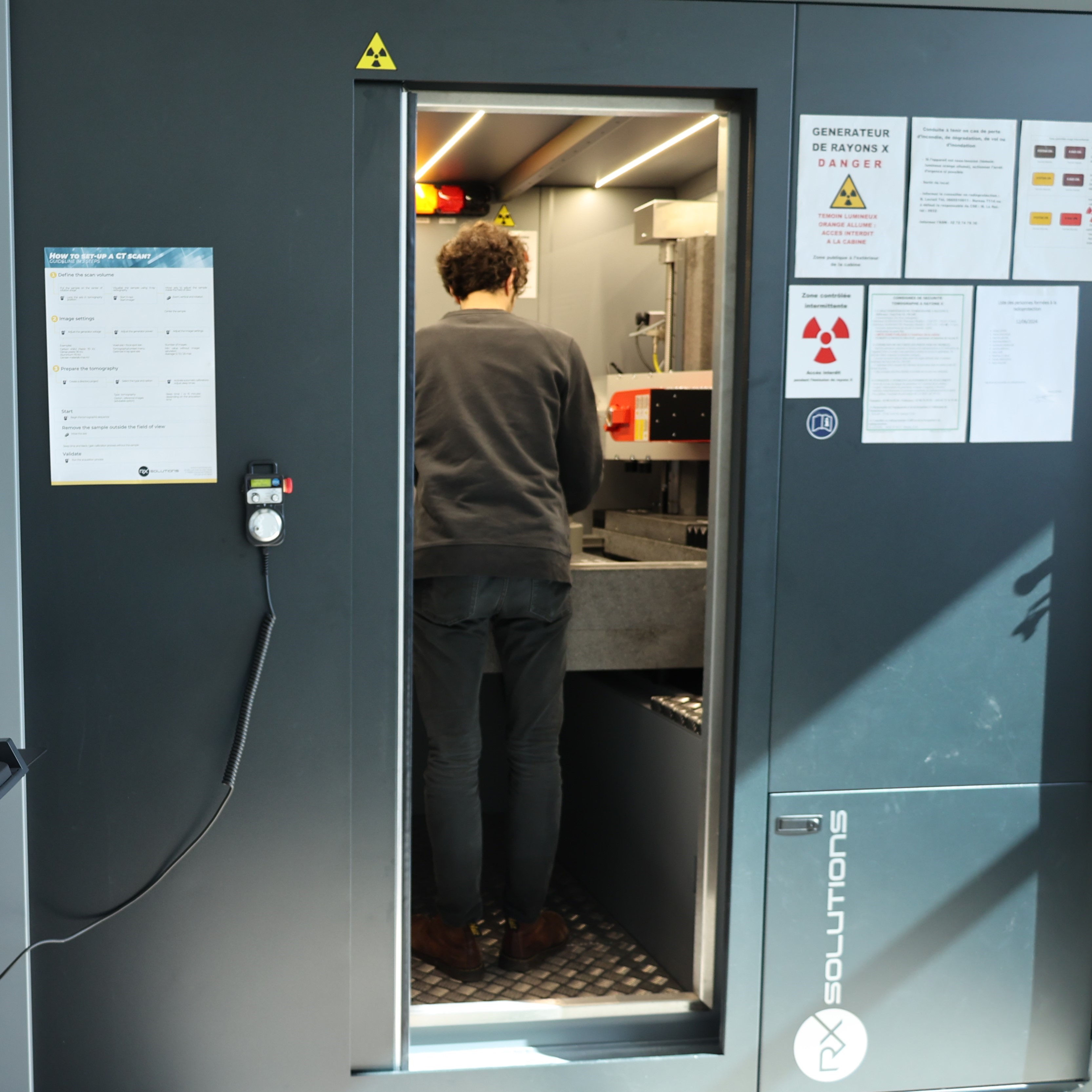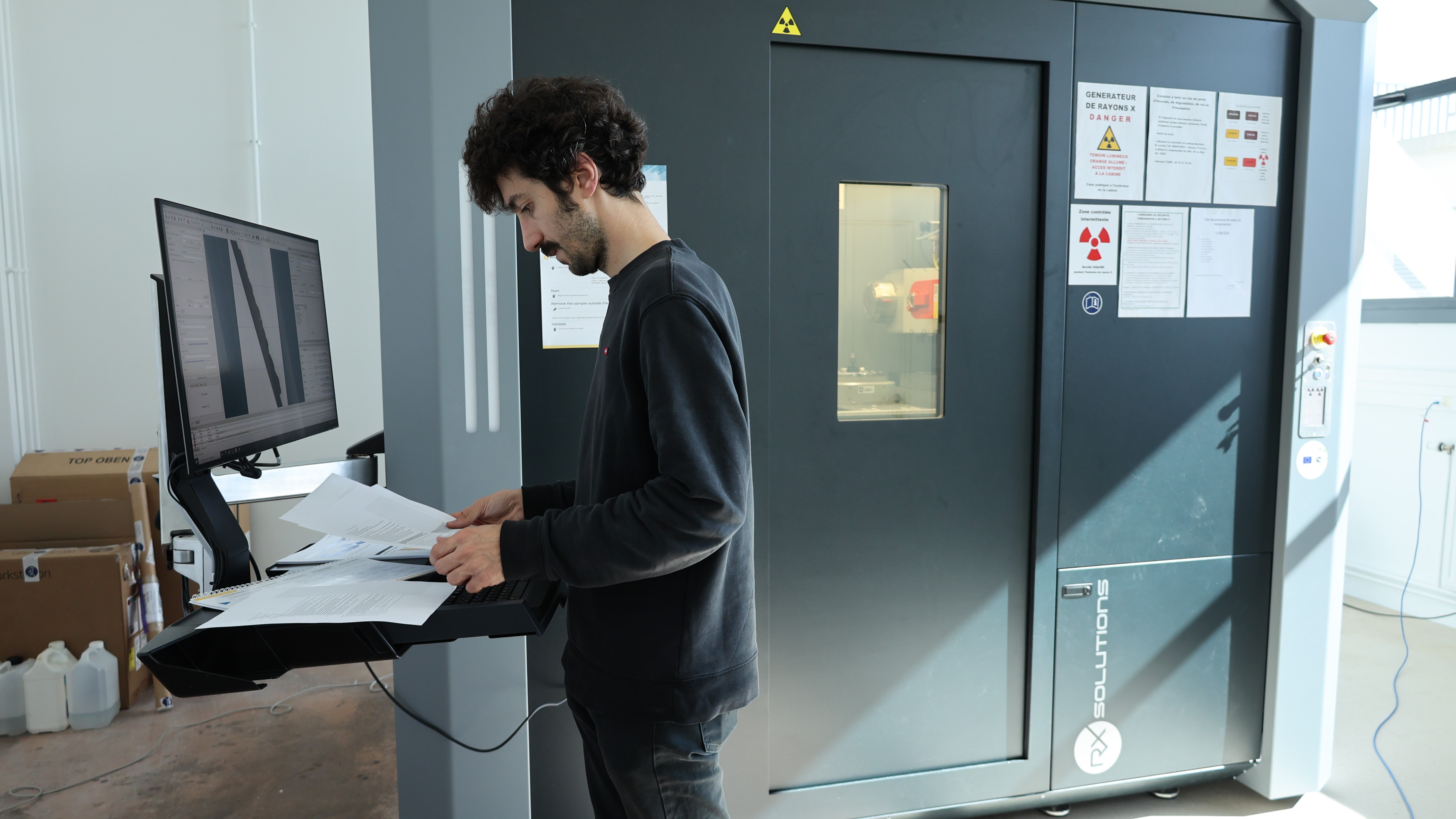

High-precision equipment for characterizing a wide range of materials
The X-ray micro-tomograph can be used to define the microstructure of the analyzed sample with great precision (a few microns or even sub-microns). Imaging, for example, reveals the air bubbles that have formed in an aluminum alloy produced by additive manufacturing. We can see the structure of polyurethane foam and its many bubbles, and the wear and tear of a textile mesh that is highly stressed and used at sea…
Mechanical stresses (tension, compression and bending) can be applied in the micro-tomograph to analyze the impact of these mechanical loads on the microstructure.
Lorenzo Bercelli is a professor at the IRDL/ ENSTA Bretagne laboratory and is responsible for this equipment:
We already had test equipment for surface measurements, but the micro-tomograph enables us to go further, with finer scales and information on materials at depth. Many of the laboratory's researchers and PhD students are planning new investigations in their research work through this tool.
State-of-the-art equipment for supervised use
A team was set up to carry out the tests on this high-tech equipment. Alongside Lorenzo Bercelli, Célia Caër, Claudiu Badulescu and Matthieu Le Saux are helping professors and PhD students with their tests. They are also the radiation protection guarantors (all the steps taken to protect people from radiation).
An acquisition financed by the CPER, 2021-2027
The X-ray micro-tomograph represents a €800,000 piece of equipment. Built in France, it is financed under the 2021-2027 regional State plan contract (“Contrat Plan Etat Région“) (If-SysMer project), which brings together several institutional funders: the State, the European Union, the Brittany Region, the Finistère Department and Brest Métropole.
Other test facilities will be added to this acquisition in the coming months.


